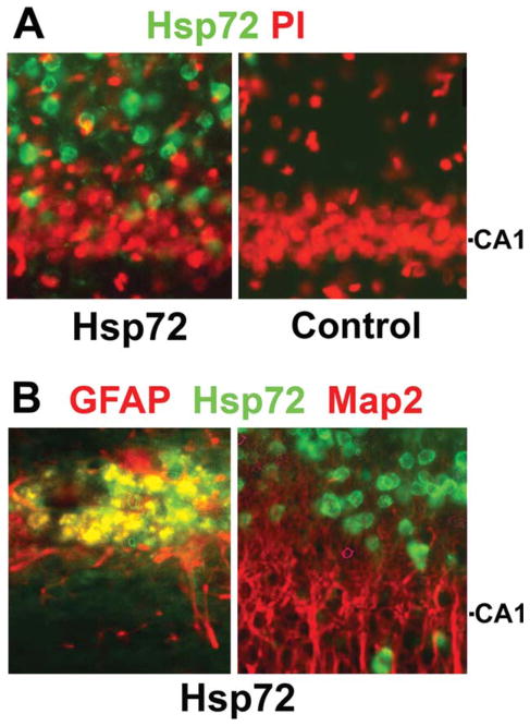
Fig. 1.
GFAPp-Hsp72 causes overexpression of Hsp72 selectively in astrocytes in vivo. Immunostained sections of brains harvested two days after stereotaxic infusion of GFAPp-Hsp72 or control DNA are shown. Sections were stained with antibody to Hsp72 (green) and then counterstained with PI (red) to show nuclei (A). The CA1 neuronal cell body level is indicated on the right. (B) Sections were double immunostained to identify astrocytes (GFAP in red, left panel) or neurons (Map2 in red, right panel). Colocalization was observed with GFAP but not Map2. Not all astrocytes were transfected, as cells staining for GFAP but not Hsp72 can be observed.

