
Fig. 5.
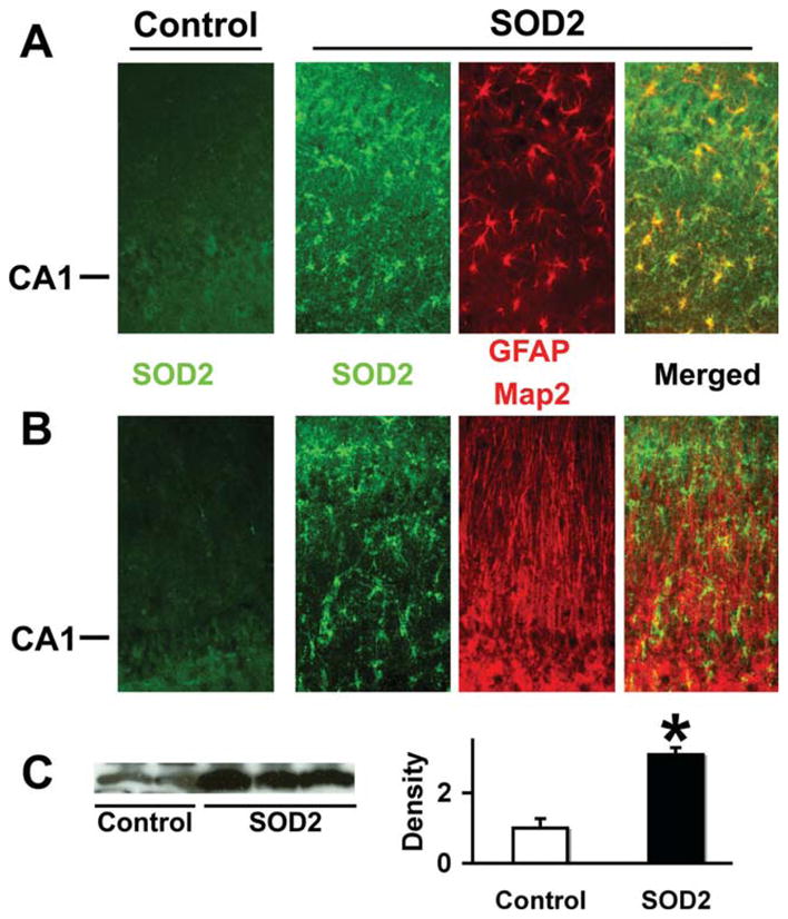
SOD2 overexpression colocalizes with the astrocyte marker GFAP but not Map2. Immunostaining of control DNA injected hippocampi demonstrated low levels of SOD2 staining (left -Control panels in A and B). SOD2 staining was significantly increased in CA1 astrocytes after stereotactic injection of GFAPp-SOD2. Double immunostaining showed SOD2 colocalized with the astrocyte marker GFAP (A) but not with neuron marker Map2 (B). The CA1 neuronal cell body layer is indicated at left. C: Western blot shows increased SOD2 protein levels in injected hippocampi compared with control plasmid injected hippocampi by densitometry (n = 3; P < 0.05).