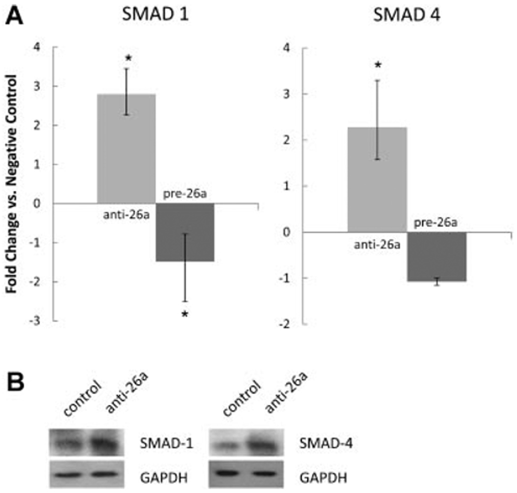
Fig. 5.
A: SMAD-1 and SMAD-4 are putative targets of miRNA-26a. Human aortic SMCs deficient in miRNA-26a display enhanced expression ofSMAD-1 and SMAD-4, while overexpression of miRNA-26a lowers expression ofSMAD-1 (*P < 0.05). Error bars indicate fold-change range derived per protocol (Applied Biosystems). B: Representative Western blots confirm increased SMAD-1/4 levels with inhibition of miRNA-26a in cells treated with 5 ng/ml of TGF-β. Left lanes: negative control antagomiR. Right lanes: anti-miRNA-26a.

