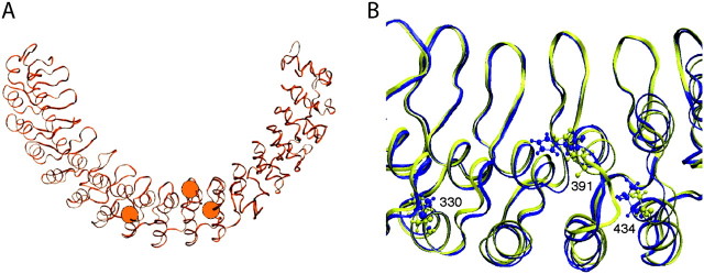
FIG. 3.
The molecular structures of ankyrin receptor. (A) The entire polyankyrin of the snake ancestor; the three orange balls illustrate amino acid sites 330, 391, and 434 (from left to right). (B) Superimposed ANK 8–ANK 11 of the snake ancestor (yellow ribbon) and its L330M/Q391H/S434T mutant (blue ribbon). The candidate amino acids at critical sites are shown with ball and stick models.

