Fig. 2.
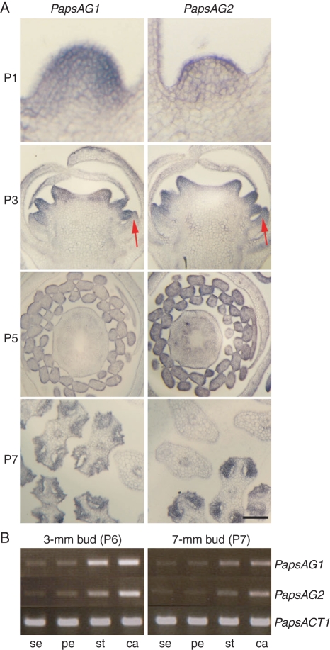
Expression analyses of PapsAG-1 and PapsAG-2. (A) In situ hybridization of PapsAG-1 and PapsAG-2 transcript-specific probes on young P. somniferum flowers showing similar expression patterns. P1 stage, Young meristem in longitudinal section before visible organ primordia appear; P3 stage, longitudinal section of developing flower bud; P5 stage, cross-section of older flower bud; P7 stage, cross-section through developing anthers with adjacent filament sections. The stages are as in Drea et al. (2007). Scale bar = 200 µm. (B) RT-PCR with PapsAG-1 and PapsAG-2 transcript-specific primers using cDNA from dissected floral organs (sepal, se; petal, pe; stamen, st; carpel, ca) of older bud stages (P6, 3-mm buds; P7, 7-mm buds). Amplification of the P. somniferum ACTIN gene, PapsACT1, was used as a control.

