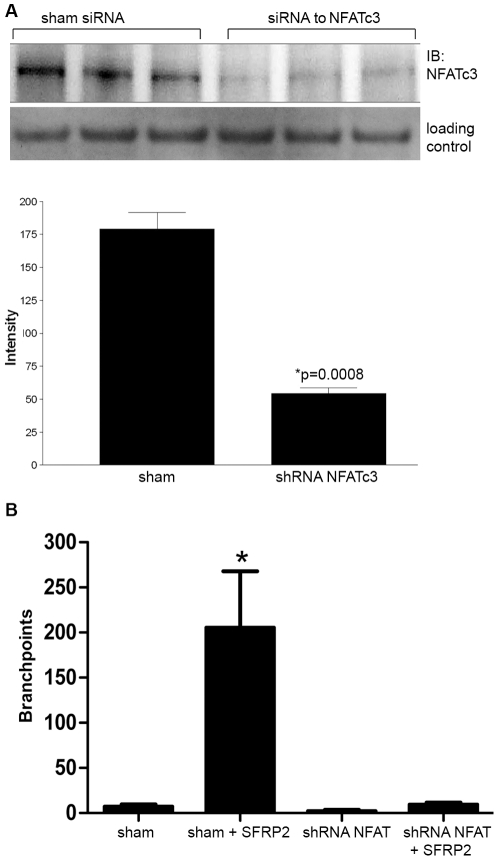
Figure 4. NFATc3 was required for SFRP2 stimulated tube formation.
A) ShRNA to SFRP2 in 2H11 endothelial cells showed 69% reduction in NFATc3 level by Western blot. The loading control was TATA binding protein TBP antibodies (a nuclear marker). B) Sham transfected 2H11 cells increased tube formation in response to SFRP2 (7 nM) (n = 3 for all groups, p<0.01), which was not seen in shRNA to NFATc3 transfected cells. Full-length blots/gels are presented in Supplemental Figure S2B. Pictures of sham transfected cells and shRNA to NFATc3 transfected cells (both stimulated with SFRP2 (7 nM)) are in Figure S3.

