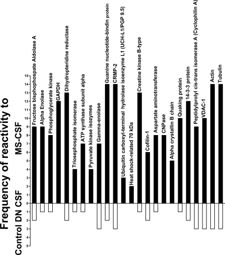
Fig. 3.
Frequency of reactivity of CSF samples to the proteins identified by mass spectrometry. This graph shows the total number of times the five MS-CSF and the control diabetic demyelinating neuropathy (DN) samples reacted to the proteins for each of the gels shown in Fig. 2 and supplemental Fig. 4. None of the other controls showed any reactivity to these proteins. Protein spots that were reactive to two or more MS-CSF samples were selected for identification by mass spectrometry and correspond to the proteins listed in Table II. Note the consistancy of the reactivity to most proteins, especially dihydropteridine reductase, guanine nucleotide-binding protein, CRMP-2, creatine kinase, actin and tubulin.

