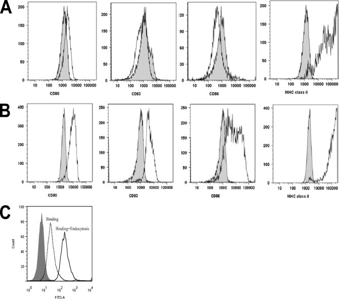
Fig. 1.
Cell surface markers of immature and mature moDCs. Cells were analyzed at the immature (A) and mature (B) state for the presence of cell surface markers CD80, CD83, CD86, and MHC class II. Gray histograms indicate isotype controls. A representative culture is depicted. C, Endocytosis and presentation of FVIII by moDCs. Histogram shows the fluorescence intensity for a FITC-labeled anti FVIII antibody (CLB-CAg-117). Binding of CLB-CAg-117-FITC to untreated cells is shown in gray. Cells stained in the absence of saponin show the membrane binding of FVIII (dotted curve). Increase of fluorescence in cells stained in the presence of saponin indicates that endocytosis of FVIII has taken place. A representative graph of one of the four donors is depicted.

