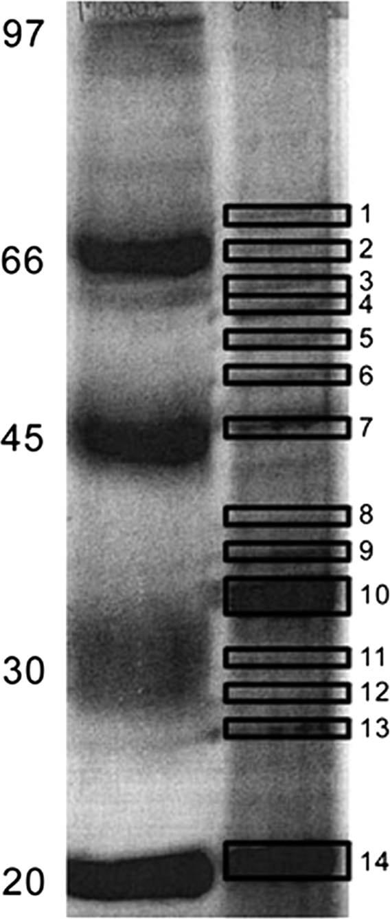
Fig. 2.

A representative gel picture of one-dimensional gel with 1 mm thickness, 12% SDS-PAGE of the membrane proteins of the Antarctic bacterium P. syringae prepared by sucrose density gradient method. 150 μg of the protein was loaded on the gel and stained the gel with coomassie blue.
