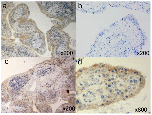
Figure 3. Immunohistochemistry of the MBP protein in human synovial tissues stained by monoclonal anti-MBP antibody.
Synovial tissue of RA patients, in particular, along the synovial lining layer strongly expressed MBP (A), whereas that of non-inflammatory osteoarthritis patients was much weaker (B). The expression of MBP in the synovial lining layer was weaker near follicules of infiltrated lymphocytes (C). Localized expression of MBP was observed at the plasma membrane of synoviocytes (D).

