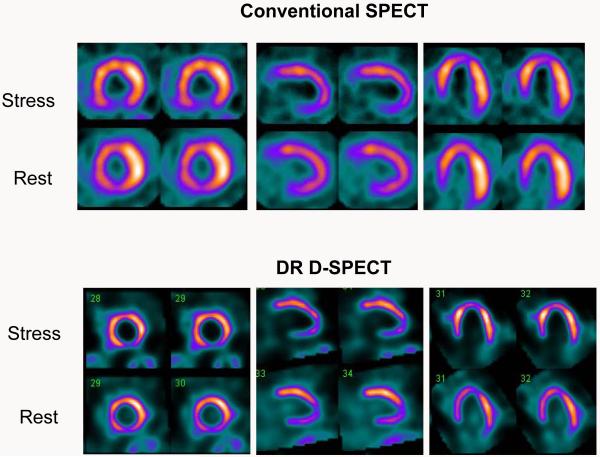
Figure 3.
66 year old male with atypical angina and shortness of breath. Known occluded RCA. Conventional SPECT images (top) showed absent uptake in the basal inferior wall and a stress induced infero-apical defect. Rest images showed infero-apical improvement. DR D-SPECT (bottom) showed similar findings.
RCA = right coronary artery; DR = dual radionuclide

