
Figure 1.
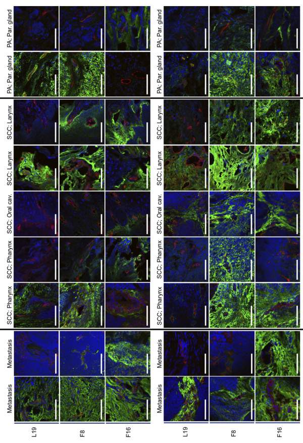
Immunofluorescence staining of head and neck cancer samples. The expression patterns and intensities of the splice isoforms extra domain A (EDA) and extradomain B (EDB) of fibronectin and the A1 domain of tenascin C were detected with the antibodies F8, L19 and F16, respectively (shown in green). A co-staining with an anti-von Willebrand factor antibody that stains blood vessels (red) and Dapi (blue) was performed. Abbreviations: SCC: squamous cell carcinoma; PA: pleomorphic adenoma.