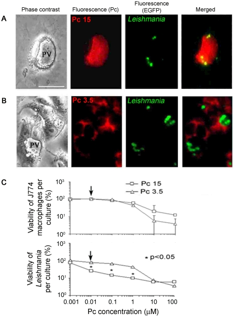
Figure 5. Exposure of infected macrophages to endocytic and mitochondrial csPcs differentially sensitize phagolysosomal GFP-Leishmania for photolysis.
[A–B] csPcs localization by fluorescence microscopy. PV (Phase contrast), Large Leishmania-containing phagolysosomes after infection of MCs with GFP-transfectants. Exposure of these cells to csPcs 15 and 3.5 (10 µM) resulted in their fluorescence at different sites: [A](Pc), csPc 15 in the PV; [A](EGFP), GFP-fluorescent Leishmania in PV; [A]Merged, Co-localization of the two in the same PV; [B](Pc), csPc 3.5 in the cytoplasm outside of PV; [B](EGFP), GFP-Leishmania in PV; [B] Merged, No co-localization of the two. Scale bar: 100 µm. [C] Viability of infected macrophages and their intracellular Leishmania . Adherent Leishmania-infected MCs as described above were exposed to increasing concentration of Pc 3.5 and Pc 15, washed and light-exposed. Upper panel, MTT assays for MC viability after 16 hrs at 35°C. Lower panel, MTT assays for viability of intracellular Leishmania released from PT-treated MCs and cultured for 7 days. □ & Δ, Samples exposed to Pc 15 and Pc 3.5, respectively. Arrows, csPc 15 versus csPc 3.5 at 0.01 µM for photolysis of intracellular Leishmania without affecting host cell viability. *, p<0.05.

