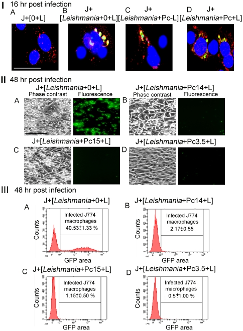
Figure 7. Infection of J774 macrophages with csPc-preloaded/pre-illuminated GFP-Leishmania, and their photolytic clearance.
[I] Endocytosis of Pc-preloaded/pre-illuminated GFP- Leishmania by J774 macrophages. [A] MCs light-exposed (J[+0+L]); [B] As [A], but pre-infected with GFP-Leishmania (J+[Leishmania+0+L]); [C] As [B], but infected with Pc 14-preloaded Leishmania without light-exposure; ([Leishmania+Pc−L]); and [D] As [C], but infected with Pc-preloaded/pre-illuminated GFP-Leishmania (J+[Leishmania+Pc+L]). Immunofluorescence microscopy of all cells 16 hr post-infection for EEA-1 endosome marker. Green, GFP-Leishmania; Blue, DAPI-stained MC nuclei; Red, EEA1-positive endosomes. Note: co-localization of Leishmania GFP with endosome marker. Scale bar: 100 µm. [II] Photolytic clearance of Pc-preloaded/pre-illuminated GFP- Leishmania from infected cells. MCs were infected with GFP-Leishmania ([A]), and those preloaded with the 3 csPcs as indicated ([B–D]) and light-exposed. Phase contrast and fluorescence microscopy of infection after 2 days. Note: Clearance of GFP from all doubly treated cultures without affecting the appearance of host cell monolayers. Scale bar: 300 µm. [III] Flow-cytometric quantitation of GFP fluorescence of the same samples used for [II], showing a ∼40% infection rate in the control ([A] GFP) reduced to negligible levels in the doubly treated groups ([B–D] GFP).

