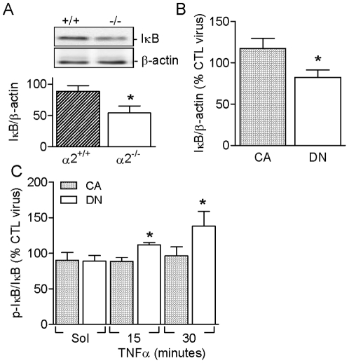
Figure 5. Role of AMPKα2 in regulating the expression and phosphorylation of IκB.
Expression of IκB in (A) pulmonary endothelial cells from AMPKα2+/+ and AMPKα2-/- mice, and (B) in COS-7 cells expressing either constitutively active (CA) or dominant negative (DN) AMPKα2. (C) IκB phosphorylation in CA- or DN-AMPKα2 expressing COS-7 cells and stimulated with solvent (Sol) or TNF-α (10 ng/mL). Data are expressed relative to values obtained in control virus-infected cells. The bar graphs summarize the results of 4 to 5 independent experiments; *P<0.05 versus CA or +/+.

