Figure 1.
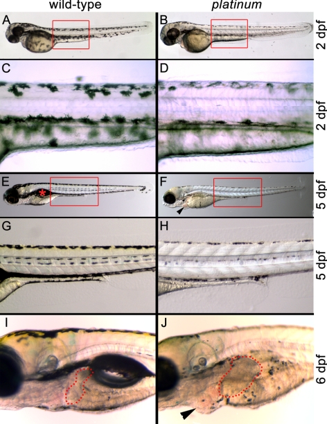
Wild-type (left) and platinum siblings (right) at 2, 5, and 6 dpf. (A) A wild-type zebrafish embryo at 2 dpf with characteristic pigmentation. Red box: the region in (C). (B) A platinum mutant embryo at 2 dpf with reduced pigmentation. Red box: region shown in (D). (C) Darkly pigmented melanophores in the wild-type embryo at 2 dpf. (D) Reduced pigmentation within each melanophore in platinum mutants at 2 dpf. (E) A wild-type zebrafish larva at 5 dpf with characteristic pigmentation and swim bladder ( ). Red box: the region in (G). (F) A platinum mutant larva at 5 dpf showing reduced pigmentation and pericardial edema (arrowhead). Red box: the region in (H). (G). Normal pigmentation pattern of a wild-type larva at 5 dpf. (H) Small melanophores with reduced pigmentation in platinum mutants at 5 dpf. (I) Location and size of the wild-type liver (outlined in red) at 6 dpf. (J) A platinum mutant at 6 dpf showing reduced pigmentation, hepatomegaly (outlined in red), and pericardial edema (arrowhead).

