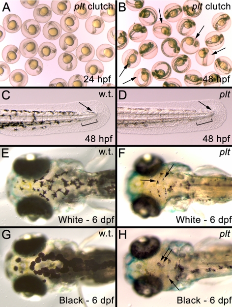
Figure 2.
(A) Image of embryos from a cross of two platinum carriers at 24 hpf. (B) The same clutch shown in A at 48 hpf. The platinum mutants (arrows) can now be distinguished from their wild-type siblings. (C) The developing tail bud of a wild-type embryo at 48 hpf showing that melanophores have migrated to the posterior of the animal (arrow) and left a gap in the ventral stripe for the developing caudal fin to appear (black bar). (D) The developing tail bud of a platinum embryo at 48 hpf showing normal melanophores migration to the tip of the tail bud (arrow) and the gap left in the ventral stripe for the developing caudal fin (black bar). (E, F) Melanophore aggregation in a wild-type larva (E) and platinum mutant (F, arrows), after 24 hours in a white environment. (G, H). Melanophore dispersion in a wild-type larva (G) and platinum mutant (H, arrows), after 24 hours in a black environment.

