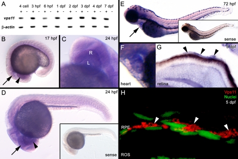
Figure 5.
(A) Temporal expression pattern of vps11 and β-actin (control) at multiple time points. cDNA reactions contained (+) or excluded (−) reverse transcriptase. (B–G) Spatial expression of vps11 as detected by whole-mount in situ hybridization. (B) A 17 hpf embryo showing ubiquitous vps11 expression, with strong expression in the developing eye field (arrowhead) and brain (arrow). (C, D) A 24-hpf embryo, showing vps11 expression in the developing retina (R, arrowhead), lens (L), and brain (arrow). (D, inset) shows the sense strand control embryo. (E) A 72-hpf embryo. Arrow: strong expression in the heart. Inset: the sense strand control embryo. (F) Expression in the heart at 72 hpf. (G) A retinal section from a 5 dpf embryo showing vps11 expression in the retina and RPE (arrowheads). (H) A retinal section showing immunolocalization of Vps11 (red, arrowheads) in the RPE at 5 dpf. RPE nuclei are stained with TO-PRO-3 (green, nuclei).

