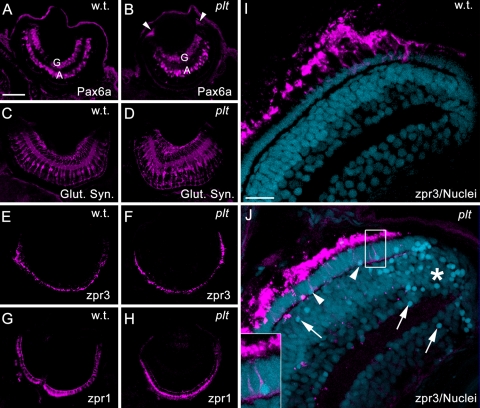
Figure 6.
(A, B) Immunolocalization of Pax6a in ganglion (G) and amacrine cells (A). The platinum mutant retinas also exhibited Pax6a expression in the retinal margins (B, arrowheads). (C, D) Immunolocalization of glutamine synthetase (Glut. Syn.) in Müller glial cells, showing hypertrophied Müller glia in the platinum mutant retinas (D). (E, F) Immunolocalization of zpr3 in rod photoreceptor outer segments. (G, H) Immunolocalization of zpr1 in double cone photoreceptors. (I, J) Immunolocalization of zpr3 (magenta) with TO-PRO-3 nuclear stain (blue). (I) A wild-type retina showing healthy rod outer segments (magenta) and uniform staining of nuclei. (J) A platinum mutant retina. Note the collapsed rod outer segments and pyknotic nuclei (arrows and arrowheads), concentrated primarily near the margins (asterisk) and in the photoreceptor layer (arrowheads). Inset: a pyknotic rod photoreceptor nucleus. Scale bar, 50 μm (A–H); 15 μm (I, J).

