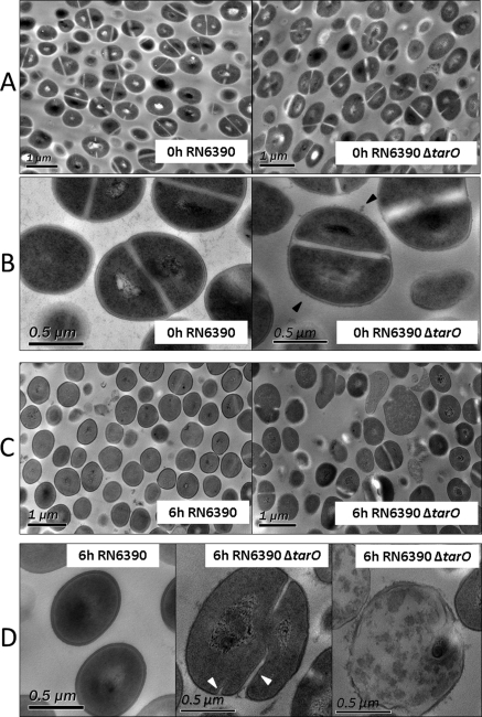
Figure 4.
Cell morphology in VH. TEM images of RN6390 and RN6390ΔtarO in VH at 0 (A, B) and 6 (C, D) hours at a lower magnification (A, C) and a higher magnification (B, D). Black arrowheads: rough surface with many surface protrusions. White arrowheads: initiation of parallel septa observed in the WTA-null mutant, RN6390ΔtarO, in VH at 6 hours.

