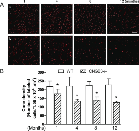
Figure 2.
Early-onset, slow progression of cone degeneration in CNGB3−/− mice evaluated by immunofluorescence labeling of S-opsin on retinal whole mounts. Immunofluorescence labeling was performed on retinal whole mounts prepared from CNGB3−/− and WT mice at 1, 4, 8, and 12 months. (A) Representative confocal images of S-opsin labeling in the inferior quadrants of the retinal whole mounts prepared from WT (a) and CNGB3−/− (b) mice. Scale bar, 10 μm. (B) Quantitative analysis of S-opsin labeling in the inferior quadrants of the retinal whole mounts. Data represent mean ± SD (n = 4–9 mice for each group). Unpaired Student's t-test was used to determine significance between age-matched CNGB3−/− and WT mice (*P < 0.05).

