Figure 5.
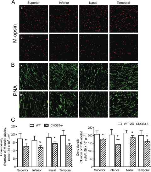
No topographic pattern of cone degeneration was found in CNGB3−/− mice. Immunofluorescence labeling for M-opsin and PNA staining was performed on retinal whole mounts prepared from CNGB3−/− and WT mice at 8 months. (A, B) Shown are confocal images of M-opsin labeling (A) and PNA labeling (B) on the quadrants of the retinal whole mounts prepared from WT (a) and CNGB3−/− mice (b). Scale bar, 10 μm. (C) Quantitative analysis of M-opsin labeling (left) and PNA labeling (right) on the quadrants of the retinal whole mounts. Data represent mean ± SD (n = 4 mice for each group). Unpaired Student's t-test was used to determine significance between age-matched CNGB3−/− and WT mice (*P < 0.05).

