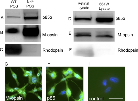
Figure 1.
p85α protein levels in cone photoreceptor outer segments and the 661W cone cell line. Western blot analysis of total mouse retinal lysates, POS-enriched extracts from WT and Nrl−/− mouse retinas, and cell extracts from the 661W cone cell line were used to assess expression levels of p85α (A, D) protein. M-cone opsin (B, E) and rhodopsin (C, F) were used as cone and rod photoreceptor markers, respectively. Immunocytochemical analysis of M-cone opsin (G) and p85α (H) expression were determined in the 661W cone cell line. For control, primary antibodies were omitted (I). Nuclei are counterstained with DAPI (blue). Scale bar, 50 μm for all panels.

