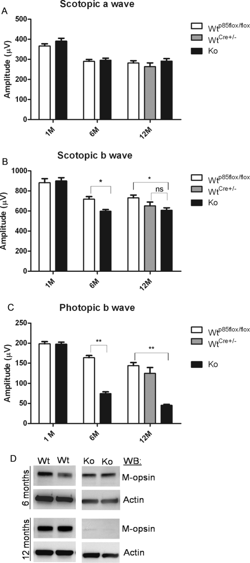
Figure 4.
Function of the cone-specific p85α KO retina. Scotopic a- and b-wave (A, B) and photopic b-wave (C) electroretinographic analysis of WT and cKO mice at 1 month (WT p85αflox/flox, n = 5; cKO, n = 5), 6 months (WT p85αflox/flox, n = 14; cKO, n = 14), and 12 months (WT p85αflox/flox, n = 10; WT Cre±, n = 4; cKO, n = 10) of age. Values are mean ± SEM. *P < 0.05 for scotopic b-wave (B) for WT p85αflox/flox vs. cKO at 6 and 12 months of age. P < 0.001 for photopic b-wave (C) for WT p85αflox/flox compared with cKO at 6 and 12 months of age. (D) Western blot analysis of WT and p85α cKO littermate retinal extracts was used to assess M-cone opsin levels at 6 and 12 months of age; β-actin was used as a loading control.

