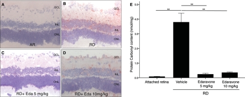
Figure 1.
Quantification of oxidative retinal damage in retina with HNE immunostaining and ELISA for PCC. Although there was minimal 4-HNE staining in AR (A), increased 4-HNE staining at the IPL and the ONL was noted 3 days after creation of RD (B). Decreased 4-HNE staining was noted after treatment with both (C) 5 (D) and 10 mg/kg edaravone. (E) There was a significant decrease in PCC 3 days after RD in the edaravone treatment group compared with the saline-treated group (P < 0.01). Data are expressed as the mean ± SE; n = 5–7. **P < 0.01. GCL, ganglion cell layer; Eda, Edaravone. Original magnification: ×200.

