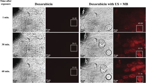
Figure 4.
Bright-field and fluorescent microscopic images of cells exposed to doxorubicin alone versus doxorubicin with ultrasound and microbubbles (US + MB). As early as 1 minute after US + MB exposures, the cells showed increased intracellular fluorescence that increased over 60 minutes. Cells exposed only to doxorubicin showed trace intracellular fluorescence at 60 minutes. Boxes represent ROIs for measuring levels of fluorescence, and values indicate mean intensity of fluorescence within the ROI.

