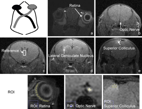
Figure 1.
T1WI of a normal mouse 1 day after topical administration of 1 M Mn2+. The Mn2+ induced signal enhancement was clearly seen in the right retina (a), right optic nerve (b), left lateral geniculate nucleus (d), and left superior colliculus (e). The ROI was selected from the retina (∼80 voxels, on the slice of the middle section of an eye), optic nerve (3 × 3 voxels square, ∼1.5 mm anterior to the chiasm), and the superior colliculis (∼25 voxels). The internal reference was a 10 × 25 rectangle in the septal area (c), which was used to normalize signals measured from the visual system.

