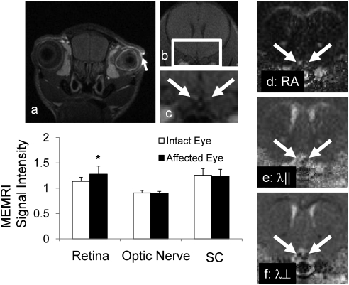
Figure 5.
MEMRI and DTI of the eye 2 weeks after retinal ischemia. (a, arrows) The remaining Mn2+ in the corneal space. There was no enhancement in the optic nerve (b, c, arrows). (c) Magnified view of the rectangle in (b). DTI characterized injured optic nerves with decreased RA (d), decreased λ‖ (e), and increased λ⊥ (f) in the affected (right) optic nerve, suggestive of the damage resulting from the retinal ischemia.

