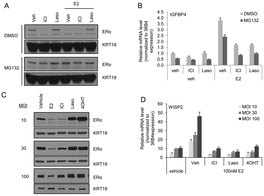
Figure 1. ICI inhibits ERα activation in despite apparent lack of ERα degradation.
A–B) MCF7 cells were treated 2 hours with DMSO or MG132 (30 µM) prior to treatment for 4 hours with vehicle (Veh), ICI 182,780 (ICI – 100 nM), or lasofoxifene (Laso – 100 nM) in the presence or absence of estradiol (E2 – 1 nM). A) Expression of ERα and loading control cytokeratin (KRT) 18 was detected by immunoblot analysis of whole cell extracts (WCE). B) ERα activation of target gene IGFBP4 was analyzed by real time quantitative PCR (qRT-PCR) analysis of samples treated in parallel with those in A. C–D) ER-negative HMECs were infected with an adenovirally expressed ERα followed by treatment for 24 hours with ER ligands: vehicle, E2 (100 nM), ICI (1 µM), Laso (1 µM), or 4-hydroxy tamoxifen (4OHT – 1µM). C) ERα and loading control cytokeratin (KRT) 18 levels were detected by immunoblot analysis of infected HMEC whole cell extracts. D) ERα activation of target gene WISP2 was analyzed by qRT-PCR of samples infected and treated in parallel with those in C. mRNA expression was normalized to similarly detected housekeeping gene 36B4 using the ΔΔCT method [21]. Data are representative of 3 independent experiments.

