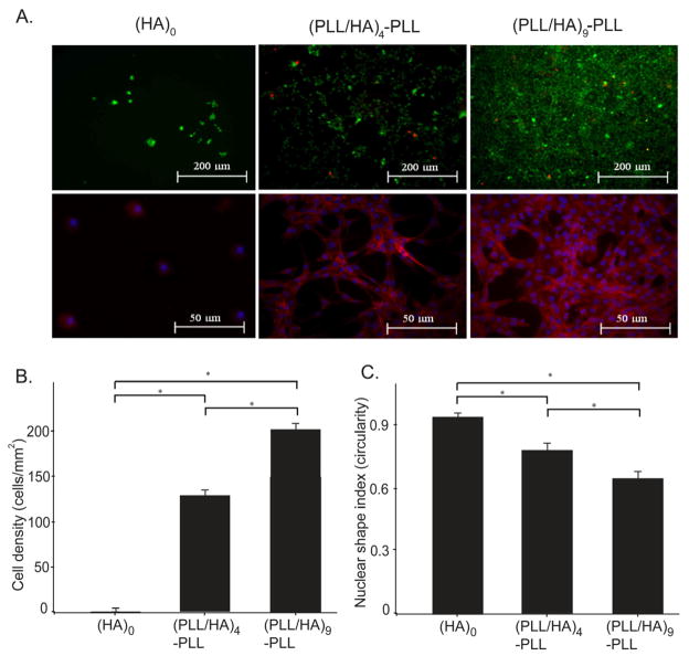
Figure 7.
NIH-3T3 cell adhesion on unmodified hydrogel (HA)0, as well as on PEM-coated hydrogels with (PLL/HA)4-PLL and (PLL/HA)9-PLL films. A. Live (green) and dead (red) as well as phalloidin staining images of NIH-3T3 cells 48 h after seeding on the hydrogel surface with and without PEM deposition. High cell viability was evidenced for cells on PEM-coated hydrogels. Phalloidin staining showed the ability of cells to adhere and spread on the hydrogel only after PEM surface modifications. B. The addition of layers significantly increased cell density (ANOVA p<0.05) as defined as the number of DAPI stained nuclei per hydrogel area. C. Layer deposition significantly increased cell spreading (ANOVA, p<0.05) as characterized by the shape index; Circularity (4*π*area/perimeter2) of each individual cell was evaluated using built-in functions of NIH ImageJ software, with a shape index of 1 representing a circle (ANOVA; Fischer’s LSD; * p<0.01).

