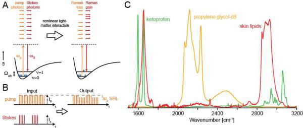
Figure 1.
Principle of SRS microscopy detection. (A) The energy diagram. During the SRS process, light is transferred from the pump beam to the Stokes beam, and the sample is vibrationally-excited. (B) Principle of modulation transfer detection. (C) Raman spectra used in this work. The contrast in SRS is based on the spontaneous Raman spectra, which are used to determine the optimal excitation wavelengths for SRS.

