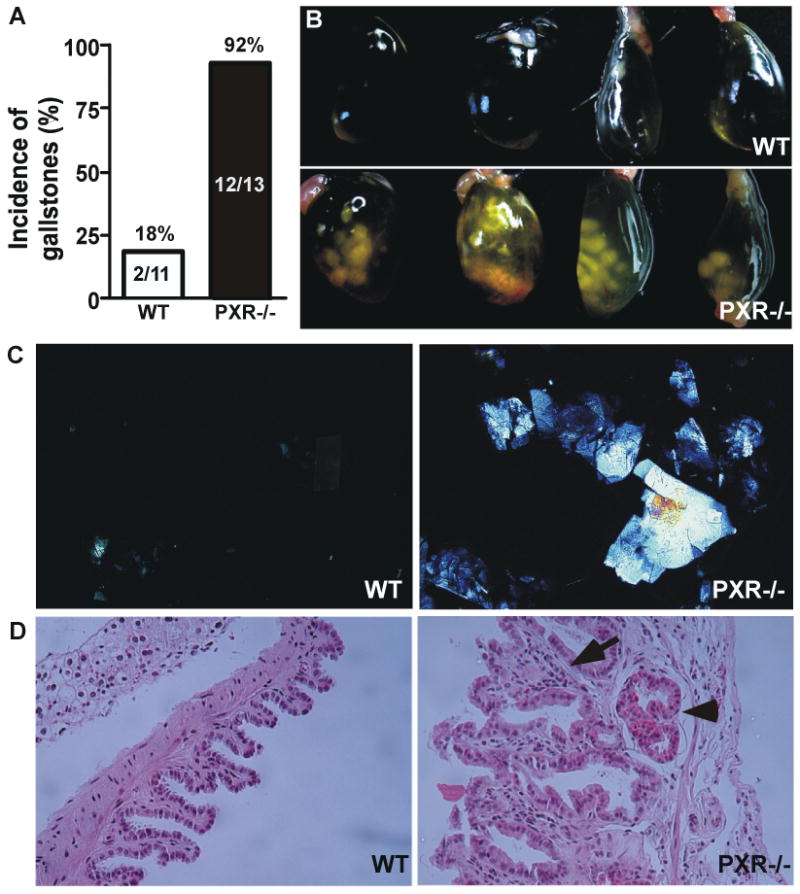
Figure 1. Loss of PXR sensitized mice to CGD.

Male WT and PXR-/- mice were subjected to lithogenic diet for 4 weeks. (A) Incidence of gallstones with the numbers of mice labeled. (B) Gross appearance of representative gallbladders. (C) Polarizing light microscopic examination of cholesterol crystals. (D) Histological examination of the gallbladder by H&E staining. The arrow indicates the thickness of the gallbladder wall and stromal granulocyte infiltration. The arrowhead indicates submucosal vasodilatation.
