Figure 2.
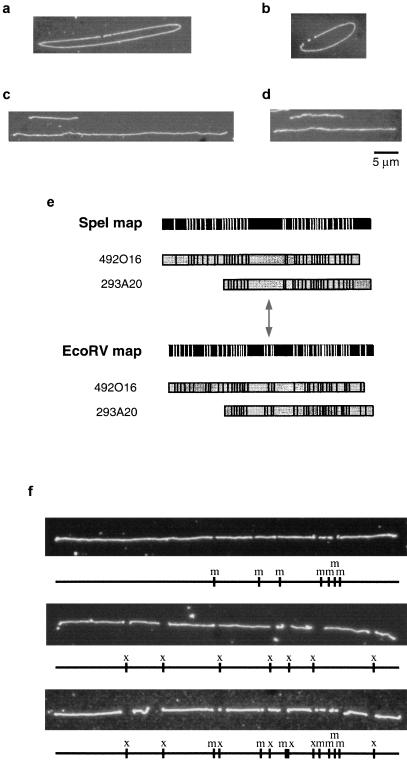
Optical mapping of BAC clones. (a,b) Digestion of BAC clones 492O16 (a) and 148I14 (b) with NotI. Small fragments represent cloning vector. Note lack of NotI sites within insert. (c,d) Sizing of BAC clones 492O16 (c) and 132B16 (d) using λ DNA as external standard. (e) EcoRV and SpeI maps of BAC clones 492O16 and 293A20. Note symmetrical arrangement of sites surrounding the small cluster of EcoRV fragments and projecting in either direction from the center of the large SpeI fragment, indicated by double-headed arrow. (f) Orientation of single-enzyme maps of clone 492K23 using double digestion approach.

