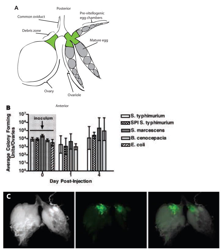
Figure 4. Bacteria colonize the fly ovary.
(a) Diagram describing the anatomy of the ovary. Each fly has two ovaries. The one on the left is shown whole while the one on the right is shown in an exploded view revealing three of the approximately 8 ovarioles that make up the ovary. Development proceeds from the anterior to the posterior end of the ovary, where eggs are laid through the common oviduct. The “debris zone” where bacteria and hemocytes are found is marked in green.
(b) Bacterial growth in ovaries of female flies injected with bacteria. n=5 ovary pairs. The geometric mean and 95% confidence intervals are indicated. See Supplemental Figure 3 for raw data and full time course.
(c) Ovaries dissected from a female fly 2 days post-injection with S. typhimurium pmig-1. DIC image (left), localization of green bacteria in the debris zone (center), overlay (right).

