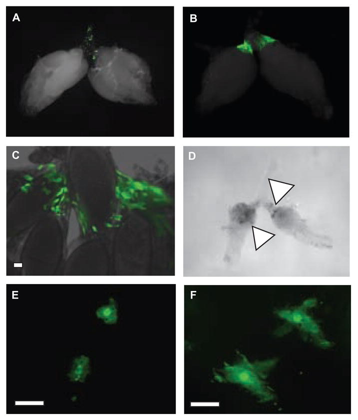
Figure 5. Fly hemocytes respond to ovary infection.

(a) Ovary dissected from a hemolectin delta-Gal4, UAS-GFP female fly 7 days post-injection with medium. Note the green fluorescent hemocytes in the debris zone.
(b–c) Ovaries dissected from hemolectin delta-Gal4, UAS-GFP female flies 7 days post-injection with S. typhimurium. The morphology of these cells differs from that seen in uninfected ovaries in (a) The white bar indicates 10 μm.
(d) DIC image of ovaries from a female fly 10 days-post injection with S. typhimurium. Note the dark regions (marked with a triangle) that appear melanized.
(e–f) Oviducts dissected from hemolectin delta-Gal4, UAS-GFP female flies 7 days post-injection with medium(e) or S. typhimurium(f). Note the diffence in the morphology of the labeled cells in uninfected versus infected ovaries. The white bar indicates 10 μm.
