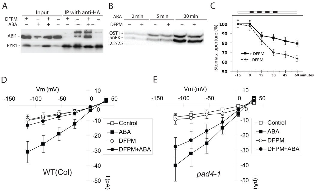
Figure 4. DFPM inhibits guard cell ABA signal transduction at the level of Ca2+ signaling.
(A) ABA-dependent protein-protein interaction between the PYR1 ABA receptor and the ABI1 PP2C-type phosphatase is not disrupted by DFPM pre-treatment. HA-PYR1 and YFP-ABI1 were co-immunoprecipitated in the presence of ABA (100 µM) and DFPM (50 µM). (B) ABA-activation (10 µM) of SnRK2 kinases, OST1, SnRK2.2, and SnRK2.3 [32] was not disrupted by DFPM treatment (50 µM). (C) DFPM (30 µM) inhibits stomatal closing mediated by repetitive imposed Ca2+-transients. Black bars represent periods in which stomata were exposed to buffer containing 1 mM CaCl2 + 1 mM KCl and white bars indicate periods with application of 0 mM CaCl2 + 50 mM KCl [43]. Each black bar corresponds to 5 minute time scale. Stomatal apertures at time = 0 (100%) correspond to average stomatal apertures of 4.02±0.25µm in control treatments and 3.53±0.26µm in DFPM pre-treatments (30 min prior to first Ca2+ pulse). Error bars show ± s.e.m (n=4 experiments). (D) ABA activation of S-type anion channel currents is significantly inhibited by DFPM in Columbia wildtype guard cells (Control: n=6; 10 µM ABA: n=10; 30 µM DFPM: n=4; 30 µM DFPM+10 µM ABA: n=10; p = 0.032, 2-tail T-test). (E) DFPM inhibition of ABA activation of S-type anion channels is not visible in pad4-1 guard cells (Control: n=6; 10 µM ABA: n=10; 30 µM DFPM: n=6; 30 µM DFPM+10 µM ABA: n=10; P = 0.314; 2-tail T-test). Guard cell protoplasts were pretreated with 0.06% DMSO (Control) or DFPM for 30 min before ABA+DMSO or ABA+DFPM treatment. Error bars show ± s.e.m.

