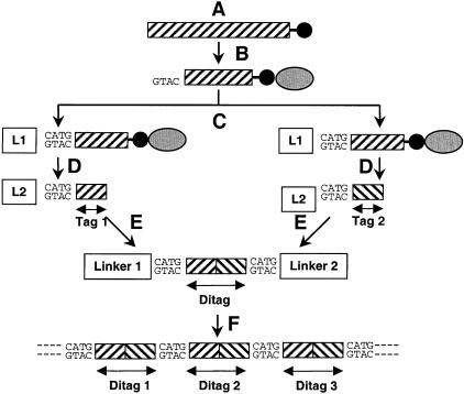
Figure 1.
Schematic illustration of the SAGE process. (A) Poly(A +)RNA is extracted and transcribed into double-stranded cDNA, primed by biotinylated oligo (dT) (black circles), and digested with the Anchoring Enzyme. (B) The 3′-most fragments are isolated by binding them to streptavidin beads (gray ellipses). (C) The fragments are divided and ligated to different linkers (L1, L2). (D) The isolated linker-tags are blunt-ended. (E) The linker-tags are ligated to linker-ditag-linker constructs and amplified by PCR (E). (F) The ditags are isolated, ligated to concatamers, cloned, and sequenced. This figure is an adaptation of Figure 1 from Velculescu, V.E. et al. (1995).

