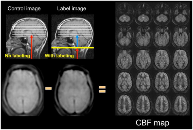
Figure 1.
ASL methodology. Images are acquired without (control) and with (label) application of arterial inversion at the site of the labeling plane. In this schematic, the inverted blood in the label image is shown in blue. Proton density images are acquired following the labeling and the post-label delay time. The label image has slightly less signal intensity due to the infusion of inverted magnetization provided by the inflowing labeled blood. When subtracted from the control image, the ASL difference images are roughly proportional to CBF. At the right are CBF images of the central 20 slices of a 32 slice whole-brain 3D-FSE imaging set acquired at 3T requiring 4:30 min to acquire.

