
Figure 10.
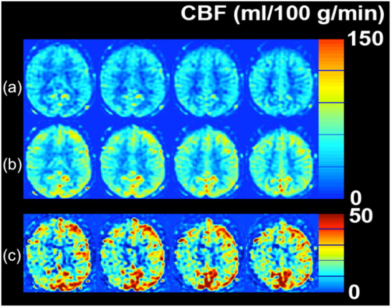
Cerebrovascular reserve (CVR) abnormality detected with MRI ASL CBF study in a 51 year-old woman with 3 hours of transient left leg weakness. ABCD2 score was 3. ASL CBF maps obtained (a) before and (b) 20 minutes following 1 g of IV acetazolamide. (c) The pre-post difference image demonstrates that both sides show CBF augmentation, with the left increasing by about 30–50 ml/100 g/min. The right hemisphere also increases, but not as much as the left, with CBF augmentation of about 10–30 ml/100 g/min. The lower CVR localizes to the expected hemisphere of the patient’s symptoms, and may put her at higher risk of future infarct in this territory.