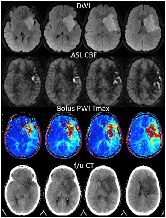
Figure 3.
25 year-old woman 6 hrs following aortic coarctation repair and inability to speak or move her right side. MR angiogram (not shown) demonstrated no flow-related enhancement in the left middle cerebral artery (MCA). Diffusion-weighted images (DWI) confirm acute ischemic stroke in the left MCA territory. ASL CBF images show hypoperfusion in the regions with high DWI signal, with arterial transit artifact (ATA) in the periphery. Bolus contrast normalized time-to-maximum (Tmax) maps show a severe abnormality in the regions corresponding to the DWI lesion, with a milder abnormality in the region of ATA on ASL. The patient received no treatment due to her recent surgery. Coregistered slices from a follow-up noncontrast CT examination 3 days later reveals that there was no increase in the size of the lesion into the region with ATA on ASL. This is consistent with prior reports suggesting that ATA may reflect the beneficial effects of collateral flow and that such tissue has a good prognosis.

