
Figure 8.
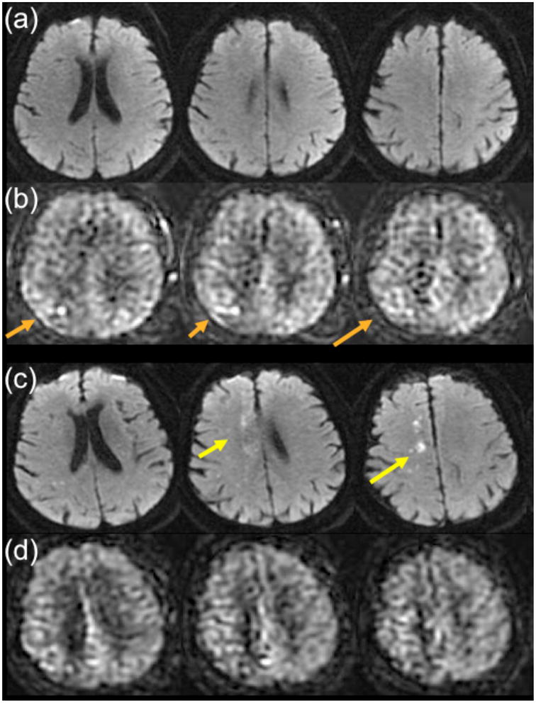
79 year-old man with transient left leg weakness. ABCD2 score was 5. (a) Initial DWI was normal. (b) ASL shows mild ATA in the right paramedian region (orange arrows) suggesting a vascular etiology for the patient’s symptoms. Two days later, the patient returned with new onset of left leg weakness and the DWI study shows new small infarcts in the right paramedian region (yellow arrows) near the prior ATA abnormality from the initial ASL. (d) ASL shows slightly more pronounced ATA in the same territory.