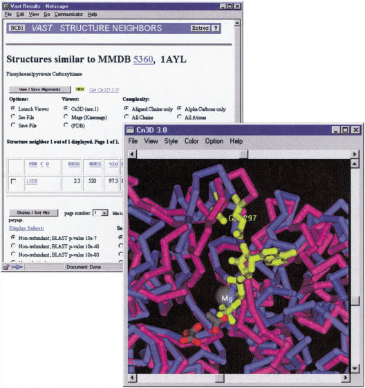
Figure 3.
Three-dimensional superposition of two PCK structures, in the presence and absence of substrates. The liganded structure 1AYL is rendered in magenta and the substrate-free structure 1OEN in blue. It can be seen that the active site cleft is closed in upon binding and that residue E297 moves into close proximity to the substrate. E297 is conserved in the protein product of APE0033 (not shown).

