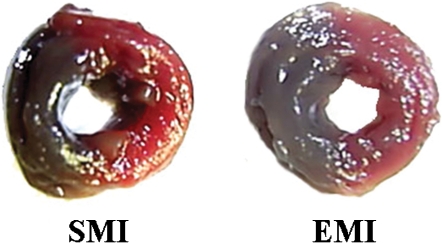
Figure 2.
Images of the midline transverse section of the heart showing the left ventricular staining by the Evans blue dye at one hour after the induction of infarction in the sedentary (SMI) and exercise (EMI) groups. The risk areas are stained red, and viable, perfused myocardium is stained blue.

