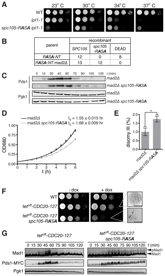
Figure 2. spc105-RASA lethality is rescued by dampened Ipl1 activity or impaired SAC.
(A) Ten-fold serial dilutions of WT, ipl1-1, and ipl1-1 spc105-RASA were plated on YEPD at 23, 30, 34, and 37°C. (B) Number of colonies with indicated genotypes or those that failed to form macroscopic colonies (DEAD) derived from single cells isolated after HGR on the strain harboring the RASA-NT cassettes in the background of wild-type or mad2Δ. (C) G1 synchronized mad2Δ and mad2Δ spc105-RASA cells were released, and Pds1 and Pgk1 (loading control) levels were monitored by Western blot. (D) Growth curve of mad2Δ and mad2Δ spc105-RASA at 30°C in YEPD medium. Average ± SEM of the doubling time of three independent experiments are also shown. (E) Disomy III formation in mad2Δ and mad2Δ spc105-RASA cells containing a chromosome III marked with a leu2 locus disrupted by URA3. The mean frequency ± SEM of disomy formation (assessed by generation Leu+, Ura+ colonies) from ten independent cultures are shown. Asterisk, p = 0.0149. (F) Ten-fold serial dilutions of WT, tetoff-CDC20-127, and tetoff-CDC20-127 spc105-RASA were plated on YEPD with or without 5 μg/ml doxycycline at 30°C. High magnification images of microcolonies are also shown. Scale bar, 50 μm. (G) G1 synchronized tetoff-CDC20-127 and tetoff-CDC20-127 spc105-RASA cells were released in the presence of doxycycline. Pds1-18MYC, Mad1, and Pgk1 (loading control) levels were analyzed by Western blots at the indicated timepoints after release. See also Figure S2.

