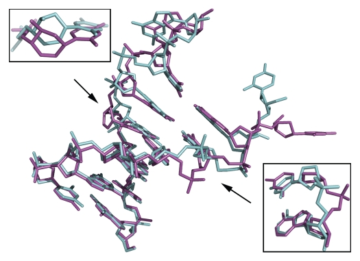
Figure 3.
Stick representation of the two CeNA substituted DNA duplexes, duplex 1 (green) and 2 (magenta), superposed onto each other. Both duplexes closely resemble each other apart from the flipped out cytosine residue. Upper left, enlargement showing the shift of the CeNA residue of duplex 2 towards the minor groove with respect to the CeNA residue in duplex 1. Lower right, enlargement showing the phosphate groups of the adenine residues, which are found at opposite sides of the sugar-phosphate backbone.

