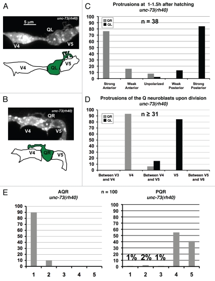
Figure 7.
UNC-73/Trio is required for robust lamellipodial protrusion. (A and B) Confocal fluorescent micrographs of Q neuroblasts in unc-73(rh40) mutants in L1 larvae visualized with scm::gfp::caax expression at 1–1.5 h after hatching. Asterisks mark the position of the Q neuroblasts. Tracings of the Q neuroblasts are located beneath each micrograph. The scale bar in (A) represents 5µm for (A and B). QL (A) and QR (B) neuroblasts polarized in the correct directions, but the sizes of the protrusions were reduced and resembling filopodial rather than robust lamellipodial protrusion seen in wild type. (C) Quantitation of the direction and extent of protrusions during the polarization stage of the Q neuroblasts at 1–1.5 h after hatching in unc-73(rh40) mutants. The graphs are organized as described in Figure 1E and Materials and Methods. For Q neuroblast polarizations, n = 38. (D) Quantitation of the location of the Q neuroblasts at division with respect to the adjacent seam cells in unc-73(rh40) mutants. The graphs are organized as described in Figure 1F and Materials and Methods. For Q neuroblast migrations, n ≥ 31. (E) Quantitation of the final migratory positions of the AQR and PQR neurons in unc-73(rh40) mutants. The graphs are organized as described in Figure 1H and Materials and Methods. For AQR and PQR migrations, n = 100.

