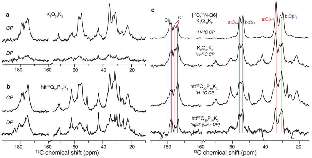
Figure 6.
PolyQ amyloid core and the effect of the httNT domain. (a) 1H-13C CP (top) and direct 13C polarization (DP; bottom) of unlabeled K2Q31K2 fibrils. These rigid sites are largely absent from the DP spectrum due to the short recycle delay (3 s). (b) Analogous spectra for unlabeled httNTQ30P10K2 fibrils indicating increased mobility in many sites. (c) Subtraction of mobile sites in the DP spectrum for httNTQ30P10K2 from its CP spectrum (3rd row) reveals the most rigid sites in this sample (bottom). These sites match the pattern of rigid polyQ signals in unlabeled K2Q31K2 fibrils (2nd row), as well as the signal from the singly labeled Gln6 in K2Q30K2 (top), which is virtually identical to Gln19 in the httNTQ30P10K2 fibril core (Figure 4). Thus, the site-specifically labeled glutamine resonances are seemingly unaffected by the presence of httNT, and appear to be representative of the bulk of the glutamines in both httNTQ30P10K2 and simple polyQ fibrils.

