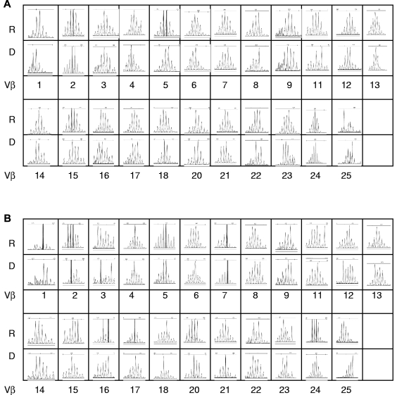
Figure 3.
Representative TCRVβ spectratype from a pair of recipient and donor pair at SCT. The spectratype profiles of the 23 Vβ subfamilies were analyzed. Shown are the CDR3 lengths on the x-axis versus the fluorescence intensity in the y-axis. R indicates recipient; D, donor. (A) CD4+ cells. (B) CD8+ cells.

