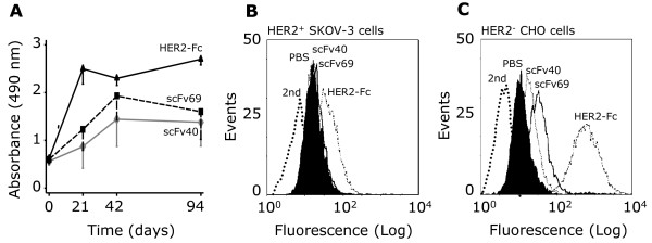
Figure 1.
Anti-HER2 antibodies are present in sera from BALB/c mice immunized with anti-Id scFv40 and scFv69. (A) ELISA of pooled sera from mice (n = 20/group) immunized with scFv40, scFv69 or HER2-Fc. Values are presented as mean ± SD of the Absorbance at 490 nm from three independent experiments. Flow cytometry analysis in (B) HER2-overexpressing SK-OV-3 and (C) HER2-negative CHO cells of the HER2-specific response of pooled sera from mice immunized with PBS, anti-Id scFv40, scFv69 or with HER2-Fc. Sera were recovered at Day 42 post-vaccination. Antibody binding was detected using a FITC-labeled goat anti-mouse antibody. FITC-labeled secondary antibody alone was used as a negative control to determine the level of background fluorescence.

