Figure 2.
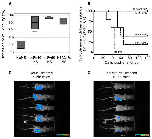
Inhibition of cell viability and analysis of SK-OV-3 tumor progression in nude mice. (A) Inhibition of cell viability in SK-OV-3 Luc cells following treatment with sera from BALB/c mice immunized with PBS (NoMS), scFv40 (scFv40MS), scFv69 (scFv69MS) or with HER2-Fc (HER2-FcMS). Cell number was proportional to the emitted luminescence and results were normalized to the fluorescence of untreated cells. Grey boxes represent the 25th and 75th percentiles with the medians as black lines; whiskers mark the smallest and largest non-outlier observations, and outliers are indicated by dots. (B) Analysis of tumor progression in nude mice (n = 5/group) xenografted with HER2-overexpressing SK-OV-3-Luc tumor cells and then adoptively transferred by i.p. injection of 200 μl of scFv40MS, scFv69MS, HER2-FcMS or NoMS (negative control), diluted 1:5, at Day 4 after xenograft. Positive controls were animals treated with trastuzumab (200 μg/injection). Tumor growth was evaluated by measuring the emitted luminescence once a week following injection of luciferin. Bioluminescence imaging of nude mice xenografted with SK-OV-3-Luc tumor cells and treated with NoMS (C) or scFv69MS (D) was performed at the end of the treatment (Day 27 after xenograft).

